

- 20 Oct
- 2025
DETEKSI ARITMIA ECG PADA DATA TIDAK SEIMBANG MENGGUNAKAN DEEP LEARNING
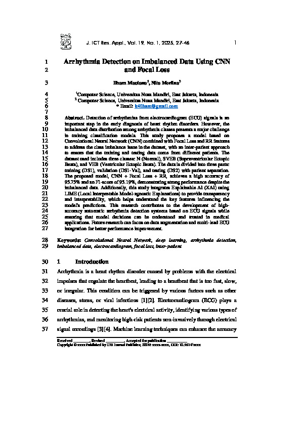
Mendeteksi aritmia dari sinyal elektrokardiogram (ECG) merupakan langkah penting dalam diagnosis dini gangguan irama jantung. Namun, distribusi data yang tidak seimbang antar kelas aritmia menjadi tantangan besar dalam pelatihan model klasifikasi. Penelitian ini mengusulkan model berbasis Convolutional Neural Network (CNN) yang dipadukan dengan Focal Loss dan fitur RR untuk mengatasi masalah ketidakseimbangan kelas dalam dataset. Dataset yang digunakan mencakup tiga kelas: N (Normal), SVEB (Supraventricular Ectopic Beats), dan VEB (Ventricular Ectopic Beats). Data dibagi menjadi tiga bagian: pelatihan (DS1), validasi (DS1-Val), dan pengujian (DS2) dengan pemisahan antar pasien. Model yang diusulkan, CNN + Focal Loss + RR, mencapai akurasi tinggi sebesar 95,75% dan F1-score sebesar 95,19%, menunjukkan performa yang kuat meskipun data yang digunakan tidak seimbang. Selain itu, penelitian ini mengintegrasikan Explainable AI (XAI) menggunakan LIME (Local Interpretable Model-agnostic Explanations) untuk memberikan transparansi dan interpretabilitas, yang membantu memahami fitur-fitur utama yang mempengaruhi prediksi model. Penelitian ini memberikan kontribusi pada pengembangan sistem deteksi aritmia otomatis berbasis sinyal ECG dengan akurasi tinggi sambil memastikan keputusan model dapat dipahami dan dipercaya dalam aplikasi medis. Penelitian selanjutnya dapat difokuskan pada augmentasi data dan integrasi multi-lead ECG untuk peningkatan kinerja yang lebih baik.
Unduhan

REFERENSI
- J. Nurriski and A. Alamsyah, “Optimasi Deep Convolutional Neural Network (Deep CNN) untuk Deteksi Aritmia Melalui Sinyal EKG Menggunakan Arsitektur Conv1D,” Indones. J. Math. Nat. Sci., vol. 46, no. 1, pp. 10–20, 2023, doi: 10.15294/ijmns.v46i1.46176.
[2] Margarita Kurti, “COVID-19: Associated subacute thyroiditis in Albania,” Int. J. Front. Life Sci. Res., vol. 2, no. 1, pp. 029–031, 2022, doi: 10.53294/ijflsr.2022.2.1.0031.
[3] Y. K. Kim, M. Lee, H. S. Song, and S. W. Lee, “Automatic Cardiac Arrhythmia Classification Using Residual Network Combined With Long Short-Term Memory,” IEEE Trans. Instrum. Meas., vol. 71, pp. 1–17, 2022, doi: 10.1109/TIM.2022.3181276.
[4] E. al. Sandhya Samant, “Exploring ECG Signal Analysis Techniques for Arrhythmia Detection: A Review,” Int. J. Recent Innov. Trends Comput. Commun., vol. 11, no. 9, pp. 4881–4896, 2023, doi: 10.17762/ijritcc.v11i9.10084.
[5] Y. Dong, M. Zhang, L. Qiu, L. Wang, and Y. Yu, “An Arrhythmia Classification Model Based on Vision Transformer with Deformable Attention,” Micromachines, vol. 14, no. 6, 2023, doi: 10.3390/mi14061155.
[6] E. Merdjanovska and A. Rashkovska, “A framework for comparative study of databases and computational methods for arrhythmia detection from single-lead ECG,” Sci. Rep., vol. 13, no. 1, pp. 1–15, 2023, doi: 10.1038/s41598-023-38532-9.
[7] M. M. Farag, “A Tiny Matched Filter-Based CNN for Inter-Patient ECG Classification and Arrhythmia Detection at the Edge,” Sensors, vol. 23, no. 3, pp. 1–23, 2023, doi: 10.3390/s23031365.
[8] X. Zhao et al., “Deep learning assessment of left ventricular hypertrophy based on electrocardiogram,” Front. Cardiovasc. Med., vol. 9, 2022, doi: 10.3389/fcvm.2022.952089.
[9] O. Ozaltin and O. Yeniay, “A novel proposed CNN–SVM architecture for ECG scalograms classification,” Soft Comput., vol. 27, no. 8, pp. 4639–4658, 2023, doi: 10.1007/s00500-022-07729-x.
[10] A. Bayani and M. Kargar, “LDCNN: A new arrhythmia detection technique with ECG signals using a linear deep convolutional neural network,” Physiol. Rep., vol. 12, no. 17, pp. 1–23, 2024, doi: 10.14814/phy2.16182.
[11] F. Kazemi Lichaee, A. Salari, J. Jalili, S. Beikmohammad Dalivand, M. Roshanfekr Rad, and M. Mojarad, “Advancements in Artificial Intelligence for ECG Signal Analysis and Arrhythmia Detection: A Review,” Int. J. Cardiovasc. Pract., vol. 8, no. 2, 2024, doi: 10.5812/intjcardiovascpract-143437.
[12] R. Holgado-Cuadrado, C. Plaza-Seco, L. Lovisolo, and M. Blanco-Velasco, “Characterization of noise in long-term ECG monitoring with machine learning based on clinical criteria,” Med. Biol. Eng. Comput., vol. 61, no. 9, pp. 2227–2240, 2023, doi: 10.1007/s11517-023-02802-5.
[13] S. Saleem, A. H. Khandoker, M. Alkhodari, L. J. Hadjileontiadis, and H. F. Jelinek, “A two-step pre-processing tool to remove Gaussian and ectopic noise for heart rate variability analysis,” Sci. Rep., vol. 12, no. 1, pp. 1–15, 2022, doi: 10.1038/s41598-022-21776-2.
[14] S. Elouaham, A. Dliou, W. Jenkal, M. Louzazni, H. Zougagh, and S. Dlimi, “Empirical Wavelet Transform Based ECG Signal Filtering Method,” J. Electr. Comput. Eng., vol. 2024, 2024, doi: 10.1155/2024/9050909.
[15] N. Sakli, C. Baccouch, H. Bellali, A. Zouinkhi, and M. Najjari, “IoT System and Deep Learning Model to Predict Cardiovascular Disease Based on ECG Signal,” Adv. Sci. Technol. Eng. Syst. J., vol. 8, no. 6, pp. 8–18, 2023, doi: 10.25046/aj080602.
[16] T. Yamane et al., “Trial of Sportswear Type ECG Sensor Device for Cardiac Safety Management during Marathon Running,” Adv. Biomed. Eng., vol. 11, pp. 151–161, 2022, doi: 10.14326/abe.11.151.
[17] B. H. Hai, “Eliminate Artifact on ECG Recording Using the Soft Threshold Setting on Wavelet Coefficients at Independent Components of ICA,” Trait. du Signal, vol. 40, no. 2, pp. 819–824, 2023, doi: 10.18280/ts.400244.
[18] J. Ding, Y. Tang, R. Chang, Y. Li, L. Zhang, and F. Yan, “Reduction in the Motion Artifacts in Noncontact ECG Measurements Using a Novel Designed Electrode Structure,” Sensors, vol. 23, no. 2, 2023, doi: 10.3390/s23020956.
[19] O. Barmak, I. Krak, S. Yakovlev, E. Manziuk, P. Radiuk, and V. Kuznetsov, “Toward explainable deep learning in healthcare through transition matrix and user-friendly features,” Front. Artif. Intell., vol. 7, no. November, pp. 1–13, 2024, doi: 10.3389/frai.2024.1482141.
[20] J. Jiang et al., “Development and Validation of a Deep-Learning Model to Detect CRP Level from the Electrocardiogram,” Front. Physiol., vol. 13, no. May, pp. 1–7, 2022, doi: 10.3389/fphys.2022.864747.
[21] S. Janbhasha and S. N. Bhavanam, “Recurrent Ascendancy Feature Subset Training Model using Deep CNN Model for ECG based Arrhythmia Classification,” Int. J. Adv. Comput. Sci. Appl., vol. 14, no. 5, pp. 639–647, 2023, doi: 10.14569/IJACSA.2023.0140568.
[22] S. Mandala, W. Jatmiko, S. Nurmaini, A. Rizal, and Adiwijaya, “OCADN: Improving Accuracy in Multi-class Arrhythmia Detection from ECG Signals with a Hyperparameter-Optimized CNN,” IEEE Access, vol. 13, no. February, pp. 34687–34705, 2025, doi: 10.1109/ACCESS.2025.3544273.
[23] H. Pham, K. Egorov, A. Kazakov, and S. Budennyy, “Machine learning-based detection of cardiovascular disease using ECG signals: performance vs. complexity,” Front. Cardiovasc. Med., vol. 10, no. July, pp. 1–11, 2023, doi: 10.3389/fcvm.2023.1229743.
[24] G. Zheng et al., “Hierarchical deep learning for autonomous multi-label arrhythmia detection and classification on real-world wearable electrocardiogram data,” Digit. Heal., vol. 10, 2024, doi: 10.1177/20552076241278942.
[25] F. Khan, X. Yu, Z. Yuan, and A. ur Rehman, “ECG classification using 1-D convolutional deep residual neural network,” PLoS One, vol. 18, no. 4 April, 2023, doi: 10.1371/journal.pone.0284791.
[26] B. M. Mathunjwa, Y. T. Lin, C. H. Lin, M. F. Abbod, M. Sadrawi, and J. S. Shieh, “ECG Recurrence Plot-Based Arrhythmia Classification Using Two-Dimensional Deep Residual CNN Features,” Sensors, vol. 22, no. 4, 2022, doi: 10.3390/s22041660.
[27] K. Sharma and R. K. Sunkaria, “Cardiac arrhythmia detection using cross-sample entropy measure based on short and long RR interval series,” J. Arrhythmia, vol. 39, no. 3, pp. 412–421, 2023, doi: 10.1002/joa3.12839.
[28] Y. Yanagisawa, W. Ibrahim, and N. Kumar, “A case of atrial fibrillation complicated by complete atrioventricular block,” SAGE Open Med. Case Reports, vol. 11, 2023, doi: 10.1177/2050313X231157486.
[29] J. Pueyo-Val et al., “Reports of Symptoms Associated with Supraventricular Arrhythmias as a Serious Adverse Drug Reaction in the Spanish Pharmacovigilance Database,” Pharmaceuticals, vol. 16, no. 8, pp. 1–12, 2023, doi: 10.3390/ph16081161.
[30] F. Hamza, “LabVIEW based ECG simulator and automatic detection system for cardiac disorders,” South Asian J. Soc. Rev., vol. 2, no. 2, pp. 1–19, 2023, doi: 10.57044/sajsr.2302.2.2.2302.
[31] J. Y. Kim et al., “The efficacy of detecting arrhythmia is higher with 7-day continuous electrocardiographic patch monitoring than with 24-h Holter monitoring,” J. Arrhythmia, vol. 39, no. 3, pp. 422–429, 2023, doi: 10.1002/joa3.12865.
[32] R. Mogili and G. Narsimha, “A novel weighted approach for automated cardiac arrhythmia beat classification using convolutional neural networks,” Int. J. Adv. Technol. Eng. Explor., vol. 9, no. 95, pp. 1508–1521, 2022, doi: 10.19101/IJATEE.2021.876071.
[33] M. S. Islam, M. N. Islam, N. Hashim, M. Rashid, B. S. Bari, and F. Al Farid, “New Hybrid Deep Learning Approach Using BiGRU-BiLSTM and Multilayered Dilated CNN to Detect Arrhythmia,” IEEE Access, vol. 10, pp. 58081–58096, 2022, doi: 10.1109/ACCESS.2022.3178710.
[34] T. I. Toma and S. Choi, “A Parallel Cross Convolutional Recurrent Neural Network for Automatic Imbalanced ECG Arrhythmia Detection with Continuous Wavelet Transform,” Sensors, vol. 22, no. 19, 2022, doi: 10.3390/s22197396.
[35] Y. Fang et al., “Electrocardiogram Signal Classification in the Diagnosis of Heart Disease Based on RBF Neural Network,” Comput. Math. Methods Med., vol. 2022, 2022, doi: 10.1155/2022/9251225.
[36] C. F. Zhao, W. Y. Yao, M. J. Yi, C. Wan, and Y. Le Tian, “Arrhythmia Classification Algorithm Based on a Two-Dimensional Image and Modified EfficientNet,” Comput. Intell. Neurosci., vol. 2022, 2022, doi: 10.1155/2022/8683855.
[37] Q. Xiao et al., “Deep Learning-Based ECG Arrhythmia Classification: A Systematic Review,” Appl. Sci., vol. 13, no. 8, 2023, doi: 10.3390/app13084964.
[38] S. Bhatia, S. K. Pandey, A. Kumar, and A. Alshuhail, “Classification of Electrocardiogram Signals Based on Hybrid Deep Learning Models,” Sustain., vol. 14, no. 24, pp. 1–15, 2022, doi: 10.3390/su142416572.
[39] J. S. Ryu et al., “Deep Learning Algorithms for Estimation of Demographic and Anthropometric Features from Electrocardiograms,” J. Clin. Med., vol. 12, no. 8, 2023, doi: 10.3390/jcm12082828.
[40] Y. Ansari, O. Mourad, K. Qaraqe, and E. Serpedin, “Deep learning for ECG Arrhythmia detection and classification: an overview of progress for period 2017–2023,” Front. Physiol., vol. 14, no. September, 2023, doi: 10.3389/fphys.2023.1246746.
[41] G. Slapničar, J. Su, and W. Wang, “Fundamental and Practical Feasibility of Electrocardiogram Reconstruction from Photoplethysmogram,” Sensors, vol. 24, no. 7, 2024, doi: 10.3390/s24072100.
[42] R. Jerath, M. Syam, and S. Ahmed, “The Future of Stress Management: Integration of Smartwatches and HRV Technology,” Sensors, vol. 23, no. 17, 2023, doi: 10.3390/s23177314.
[43] S. Chopannejad, A. Roshanpoor, and F. Sadoughi, “Attention-assisted hybrid CNN-BILSTM-BiGRU model with SMOTE–Tomek method to detect cardiac arrhythmia based on 12-lead electrocardiogram signals,” Digit. Heal., vol. 10, 2024, doi: 10.1177/20552076241234624.
[44] F. Pang, C. Lei, and J. Zeng, “Electrical insulator defect detection with incomplete annotations and imbalanced samples,” IET Gener. Transm. Distrib., vol. 18, no. 4, pp. 694–715, 2024, doi: 10.1049/gtd2.13107.
[45] R. Li et al., “GTF: An Adaptive Network Anomaly Detection Method at the Network Edge,” Secur. Commun. Networks, vol. 2021, no. Ml, 2021, doi: 10.1155/2021/3017797.
[46] A. M. El Koshiry, E. H. I. Eliwa, T. A. El-Hafeez, and M. Khairy, “Detecting cyberbullying using deep learning techniques: a pre-trained glove and focal loss technique,” PeerJ Comput. Sci., vol. 10, pp. 1–33, 2024, doi: 10.7717/peerj-cs.1961.
[47] Y. Y. Choi et al., “Diagnostic usefulness of implantable loop recorder in patients with unexplained syncope or palpitation,” Int. J. Arrhythmia, vol. 23, no. 1, 2022, doi: 10.1186/s42444-022-00068-w.
[48] Y. Ramadhan and S. Mandala, “Analysis of Electrocardiogram Dynamic Features for Arrhythmia Classification,” J. Online Inform., vol. 8, no. 2, pp. 204–212, 2023, doi: 10.15575/join.v8i2.1106.
[49] Z. Liu et al., “Multiclass Arrhythmia Detection and Classification From Photoplethysmography Signals Using a Deep Convolutional Neural Network,” J. Am. Heart Assoc., vol. 11, no. 7, 2022, doi: 10.1161/JAHA.121.023555.
[50] J. Hong, S. J. Litt, and J. P. Moak, “Cardiac arrhythmias in postural tachycardia syndrome and orthostatic intolerance,” Cardiol. Young, vol. 33, no. 2, pp. 255–259, 2023, doi: 10.1017/S1047951122000580.
[51] L. Saclova, A. Nemcova, R. Smisek, L. Smital, M. Vitek, and M. Ronzhina, “Reliable P wave detection in pathological ECG signals,” Sci. Rep., vol. 12, no. 1, pp. 1–14, 2022, doi: 10.1038/s41598-022-10656-4.
[52] M. Samir, H. Darwis, F. Umar, and P. Korespondensi, “Fourier Descriptor Pada Klasifikasi Daun Herbal Menggunakan Support Vector Machine Dan Naive Bayes Fourier Descriptor on Classification of Herbal Leaves Using Support Vector Machine and Naive Bayes,” vol. 10, no. 6, pp. 1205–1212, 2023, doi: 10.25126/jtiik.2023107309.
[53] M. I. Nugraha and A. Armansyah, “Penutupan Kompetensi Keahlian SMK dengan Pendekatan Klasifikasi Minat Siswa Menggunakan Jaringan Syaraf Tiruan,” J. Teknol. Sist. Inf. dan Apl., vol. 6, no. 3, pp. 269–275, 2023, doi: 10.32493/jtsi.v6i3.30143.
[54] D. Safitri, Susanti, Rahmaddeni, and T. A. Fitri, “Perbandingan Algoritma XGBoost dan SVM Dalam Analisis Opini Publik Pemilihan Presiden 2024,” Indones. J. Comput. Sci., vol. 13, no. 3, pp. 4763–4775, 2024, doi: 10.33022/ijcs.v13i3.4041.
[55] R. M. Dhodapkar, E. Li, K. Nwanyanwu, R. Adelman, S. Krishnaswamy, and J. C. Wang, “Deep learning for quality assessment of optical coherence tomography angiography images,” Sci. Rep., vol. 12, no. 1, pp. 1–9, 2022, doi: 10.1038/s41598-022-17709-8.
[56] S. S. KABIR, M. S. AHMMED, M. M. SIDDIQUE, R. R. EMA, M. RAHMAN, and S. Md. GALIB, “Breast Tumor Prediction and Feature Importance Score Finding Using Machine Learning Algorithms,” Radioelectron. Comput. Syst., vol. 4225, no. 4, pp. 32–42, 2023, doi: 10.32620/REKS.2023.4.03.
[57] E. Miranda, S. Adiarto, F. M. Bhatti, A. Y. Zakiyyah, M. Aryuni, and C. Bernando, “Understanding Arteriosclerotic Heart Disease Patients Using Electronic Health Records: A Machine Learning and Shapley Additive exPlanations Approach,” Healthc. Inform. Res., vol. 29, no. 3, pp. 228–238, 2023, doi: 10.4258/hir.2023.29.3.228.
[58] A. M. Majid and I. Nawangsih, “Perbandingan Metode Ensemble Untuk Meningkatkan Akurasi Algoritm Machine Learning Dalam Memprediksi Penyakit Breast Cancer (Kanker Payudara),” J. SAINTIKOM (Jurnal Sains Manaj. Inform. dan Komputer), vol. 23, no. 1, p. 97, 2024, doi: 10.53513/jis.v23i1.9563.
[59] S. - and A. -, “Klasifikasi Status Kemiskinan Menggunakan Algoritma Random Forest,” Eig. Math. J., vol. 5, no. 1, pp. 37–44, 2022, doi: 10.29303/emj.v5i1.133.
[60] N. Alamatsaz, L. Tabatabaei, M. Yazdchi, H. Payan, N. Alamatsaz, and F. Nasimi, “A lightweight hybrid CNN-LSTM explainable model for ECG-based arrhythmia detection,” Biomed. Signal Process. Control, vol. 90, pp. 1–7, 2024, doi: 10.1016/j.bspc.2023.105884.
[61] S. Chen, H. Wang, H. Zhang, C. Peng, Y. Li, and B. Wang, “A novel method of swin transformer with time-frequency characteristics for ECG-based arrhythmia detection,” Front. Cardiovasc. Med., vol. 11, no. June, pp. 1–12, 2024, doi: 10.3389/fcvm.2024.1401143.
[62] A. R. Hassan and M. I. H. Bhuiyan, “Automated identification of sleep states from EEG signals by means of ensemble empirical mode decomposition and random under sampling boosting,” Comput. Methods Programs Biomed., vol. 140, pp. 201–210, 2017, doi: 10.1016/j.cmpb.2016.12.015.
Kiriman Terbaru
-
07 Jul 2026
-

06 Jul 2026
-
02 Jul 2026
-
29 Jun 2026
-

22 Jun 2026