

- 06 Mar
- 2026
PREDIKSI DINI BATU EMPEDU BERBASIS BIOIMPEDANSI DAN DATA LABORATORIUM MENGGUNAKAN MACHINE LEARNING TEROPTIMASI
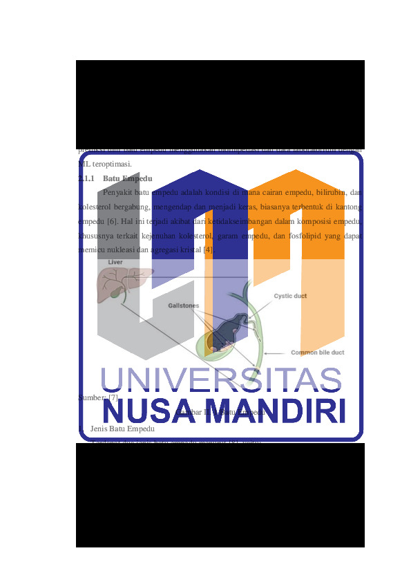
Penyakit batu empedu (kolelitiasis) merupakan gangguan gastrointestinal yang sering
bersifat asimtomatik pada tahap awal, namun berisiko menimbulkan komplikasi serius
jika tidak terdeteksi. Penelitian ini mengembangkan kerangka kerja machine learning
teroptimasi untuk prediksi dini non-invasif menggunakan data bioimpedansi dan
pemeriksaan laboratorium rutin. Dataset publik UCI Gallstone digabungkan dengan
data rumah sakit lokal dan diperkaya melalui feature engineering untuk membentuk
fitur komposit bermakna secara klinis, seperti rasio lipid, indeks komposisi tubuh, skor
komorbiditas, dan laju filtrasi glomerulus. Tujuh model klasifikasi dioptimasi
menggunakan Random Search dan dievaluasi melalui skema holdout (80:20) serta 10
fold stratified cross-validation. Hasil evaluasi menunjukkan ANN teroptimasi
mencapai akurasi 91,19%, recall 95,65%, dan ROC-AUC 0,979, sementara Random
Forest teroptimasi memperoleh AUC tertinggi 0,982. Analisis interpretabilitas
menggunakan SHAP mengidentifikasi jenis kelamin, total body water, hemoglobin,
serta rasio lipid sebagai prediktor utama. Temuan ini menunjukkan potensi pendekatan
sebagai sistem pendukung keputusan klinis yang akurat dan hemat biaya untuk
skrining dini batu empedu.
Unduhan
-
File_6 Bab V Penutup_u.pdf
Terakhir download 03 Jun 2026 14:06File_6 Bab V Penutup
- diunduh 1x | Ukuran 81 KB
-
File_3 Bab II Landasan Teori_u.pdf
Terakhir download 03 Jun 2026 14:06File_3 Bab II Landasan Teori
- diunduh 1x | Ukuran 288 KB
-
File_8 Draf Paper.pdf
Terakhir download 22 Mar 2026 02:03File_8 Draf Paper
- diunduh 4x | Ukuran 1,187 KB
-
File_2 Bab I Pendahuluan_u.pdf
Terakhir download 03 Jun 2026 14:06File_2 Bab I Pendahuluan
- diunduh 1x | Ukuran 156 KB
-
File_7 Lampiran dll_u.pdf
Terakhir download 24 Jul 2026 09:07File_7 Lampiran dll
- diunduh 85x | Ukuran 1,139,628
-
File_5 Bab IV Pembahasan_u.pdf
Terakhir download 03 Jun 2026 14:06File_5 Bab IV Pembahasan
- diunduh 2x | Ukuran 2,812,841
-
File_4 Bab III Metode Penelitian_u.pdf
Terakhir download 03 Jun 2026 14:06File_4 Bab III Metode Penelitian
- diunduh 2x | Ukuran 798,156
-
REFERENSI
[1] C. Chakraborty and N. Mukherjee, “Bayesian Hybrid Machine Learning of Gallstone Risk,” pp. 1–25, 2025, [Online]. Available: http://arxiv.org/abs/2506.14561
[2] M. W. Jones, C. B. Weir, and M. Marietta, “Gallstones (Cholelithiasis),” in StatPearls [Internet], Updated 20., Treasure Island (FL): StatPearls Publishing, 2025. [Online]. Available: https://www.ncbi.nlm.nih.gov/books/NBK459370/
[3] C. Calolus, “Batu Empedu (Tidak) Perlu Dioperasi?” Accessed: Sep. 01, 2025. [Online]. Available: https://rscarolus.or.id/artikel/batu-empedu-tidak-perlu-dioperasi/
[4] I. Esen, H. Arslan, S. Akturk Esen, M. Gulsen, N. Kultekin, and O. Ozdemir, “Early prediction of gallstone disease with a machine learning-based method from bioimpedance and laboratory data,” Med. (United States), vol. 103, no. 8, p. E37258, 2024, doi: 10.1097/MD.0000000000037258.
[5] Y. Wang et al., “Establishing a preoperative predictive model for gallbladder adenoma and cholesterol polyps based on machine learning: a multicentre retrospective study,” World J. Surg. Oncol. , vol. 23, no. 1, 2025, doi: 10.1186/s12957-025-03671-y.
[6] S. M. Abdu and E. M. Assefa, “Prevalence of gallstone disease in Africa: a systematic review and meta-analysis,” BMJ Open Gastroenterol., vol. 12, no. 1, pp. 1–8, 2025, doi: 10.1136/bmjgast-2024-001441.
[7] Y. Miyah, M. Benjelloun, H. El Omari, K. El-Mouhdi, and M. El Feniche, “Recent investigation of the medicinal plants’ effectiveness in the natural management of urinary and gallstones: a review,” Phytomedicine Plus, vol. 5, no. 3, 2025, doi: 10.1016/j.phyplu.2025.100839.
[8] M. C. Staff, “Gallstones - Symptoms & causes - Mayo Clinic.” Accessed: Sep. 04, 2025. [Online]. Available: https://www.mayoclinic.org/diseases-conditions/gallstones/symptoms-causes/syc-20354214
[9] G. P. K. Bali, B. Singh, M. Ashraf, and T. Kamalanathan, “Factors prevailing for gallstone formation,” in Gallstone Formation, Diagnosis, Treatment and Prevention, Elsevier, 2024, pp. 39–50. doi: 10.1016/B978-0-443-16098-1.00005-9.
[10] “Gallstones - NIDDK.” Accessed: Sep. 04, 2025. [Online]. Available: https://www.niddk.nih.gov/health-information/digestive-diseases/gallstones
[11] “Definition & Facts for Gallstones - NIDDK.” Accessed: Sep. 04, 2025. [Online]. Available: https://www.niddk.nih.gov/health-information/digestive-diseases/gallstones/definition-facts?
[12] R. Hasan, F. Allahbakhshi, A. D. Shlyk, and K. Allahbakhshi, “Gallstones as a predictor of elevated cardiovascular disease risk: A meta-analysis and meta-regression of over 7.4 million participants,” PLoS One, vol. 20, no. 3, p. e0314661, Mar. 2025, doi: 10.1371/journal.pone.0314661.
[13] W. Y. Dan, Y. S. Yang, L. H. Peng, G. Sun, and Z. K. Wang, “Gastrointestinal microbiome and cholelithiasis: Current status and perspectives,” World J. Gastroenterol., vol. 29, no. 10, pp. 1589–1601, 2023, doi: 10.3748/wjg.v29.i10.1589.
[14] H. Li et al., “The gut microbiota features and the application value in predicting recurrent risks for gallstone patients who underwent laparoscopic cholecystectomy,” mSystems, vol. 10, no. 8, 2025, doi: 10.1128/msystems.01760-24.
[15] Z. Wu, S. Jiang, J. Li, P. Wang, and Y. Chen, “Association between urinary cadmium levels and increased gallstone disease in US adults,” Sci. Rep., vol. 15, no. 1, pp. 1–11, 2025, doi: 10.1038/s41598-025-00648-5.
[16] J. Wang et al., “The association between blood heavy metals and gallstones: A cross-sectional study,” Sci. Total Environ., vol. 904, p. 166735, Dec. 2023, doi: 10.1016/j.scitotenv.2023.166735.
[17] Q. Zhuang et al., “Association between sleep and gallstone disease in United States adults: A cross-sectional study,” BMC Public Health, vol. 24, no. 1, p. 3291, Nov. 2024, doi: 10.1186/s12889-024-20824-y.
[18] S. Hosseini, A. Asadizeidabadi, E. Tarabrin, S. Muraviev, and D. Orlushin, “Asymptomatic Cholecystitis Presents a New Challenge for Correcting Treatment Tactics in Patients with Gallstone Disease Rather Than Being an Unsolvable Problem of Biliary Surgery,” Am. J. Intern. Med., vol. 12, no. 3, pp. 26–32, 2024, doi: 10.11648/j.ajim.20241203.11.
[19] M. S. Ul Hassan et al., “Prevalence and Associated Risk Factors of Acute Pancreatitis in Patients With Gallstones: A Cross-Sectional Study,” Cureus, May 2025, doi: 10.7759/cureus.84220.
[20] Y. Saiman, “Batu empedu - Gangguan Hati dan Kandung Empedu - Manual MSD Versi Konsumen.” Accessed: Sep. 04, 2025. [Online]. Available: https://www.msdmanuals.com/id/home/gangguan-hati-dan-kandung-empedu/gangguan-kantung-empedu-dan-saluran-empedu/batu-empedu
[21] A. R. Amaliya et al., “Komplikasi Kolesistitis : Empiema dan Hidrops Kandung Empedu,” Unram Med. J., vol. 12, no. 4, pp. 429–433, 2023, doi: 10.29303/jku.v12i4.985.
[22] M. Matan, “Analisis Bioimpedansi | Aplikasi dan Penggunaannya.” Accessed: Sep. 04, 2025. [Online]. Available: https://www.electricity-magnetism.org/id/analisis-bioimpedansi-aplikasi-dan-penggunaannya/
[23] X. Wen et al., “Clinlabomics: leveraging clinical laboratory data by data mining strategies,” BMC Bioinformatics, vol. 23, no. 1, pp. 1–20, 2022, doi: 10.1186/s12859-022-04926-1.
[24] I. S. Maesaroh, K. N. Syaja’Ah, Y. S. Perkasa, S. Hadianti, D. Riana, and R. R. Nurmalasari, “Cervical Cancer Classification From Pap Smear Using XCeption Model,” in 2024 10th International Conference on Wireless and Telematics (ICWT), IEEE, Jul. 2024, pp. 1–5. doi: 10.1109/ICWT62080.2024.10674721.
[25] H. D. Saputra, A. I. E. Efendi, E. Rudini, D. Riana, and A. S. Hewiz, “Segmentation in Identifying the Development of Ground Glass Opacity on CT-Scan Images of the Lungs,” J. Med. Informatics Technol., pp. 1–6, 2023, doi: 10.37034/medinftech.v1i1.1.
[26] W. Dirsam and M. D. Anasanti, “Improving Obesity Classification with Advanced Machine Learning and Feature Selection Methods,” Int. J. Intell. Eng. Syst., vol. 18, no. 7, pp. 373–387, Aug. 2025, doi: 10.22266/ijies2025.0831.26.
[27] K. Kirso and M. D. Anasanti, “Telematika Improving Alzheimer ’ s Disease Prediction Accuracy using Feature Selection , K Fold Cross Validation , and KNN Imputer Techniques,” Telematika, vol. 18, no. 1, pp. 75–90, 2025, [Online]. Available: https://ejournal.amikompurwokerto.ac.id/index.php/telematika/article/view/3055
[28] Mahendra and M. D. Anasanti, “Comprehensive Machine Learning Model for Cervical Cancer Prediction and Risk Factor Identification,” Hum. Behav. Emerg. Technol., vol. 2025, no. 1, 2025, doi: 10.1155/hbe2/6629232.
[29] S. Agustiani and Y. Rianto, “Deep Learning for Histopathological Image Analysis: A Convolutional Neural Network Approach to Colon Cancer Classification,” Telematika, vol. 17, no. 1, pp. 39–51, 2024, doi: https://doi.org/10.21107/rekayasa.v15i2.141580.
[30] N. Merlina, A. Prasetyo, I. Zuniarti, N. A. Mayangky, D. N. Sulistyowati, and F. Aziz, “Improving Early Detection of Cervical Cancer Through Deep Learning-Based Pap Smear Image Classification,” J. Appl. Data Sci., vol. 6, no. 2, pp. 969–980, 2025, doi: 10.47738/jads.v6i2.576.
[31] R. L. Hasanah, Y. Rianto, and D. Riana, “Identification of Acne Vulgaris Type in Facial Acne Images Using GLCM Feature Extraction and Extreme Learning Machine Algorithm,” Rekayasa, vol. 15, no. 2, pp. 204–214, 2022, doi: 10.21107/rekayasa.v15i2.14580.
[32] M. Mahendra, J. Jumadi, and D. Riana, “Cervical Cancer Papsmear Classification through Meta-Learning Technique using Convolution Neural Networks.,” J. Med. Informatics Technol., pp. 105–108, 2023, doi: 10.37034/medinftech.v1i4.23.
[33] N. Merlina, E. Noersasongko, P. N. Andono, M. Arief Soeleman, and D. Riana, “Optimization of the Preprocessing Method for Edge Detection on Overlapping Cells at PAP Smear Images,” Int. J. Informatics Vis., vol. 7, no. 2, pp. 471–476, 2023, doi: 10.30630/joiv.7.2.1329.
[34] A. M. Obaid, A. Turki, H. Bellaaj, and M. Ksantini, “Diagnosis of Gallbladder Disease Using Artificial Intelligence: A Comparative Study,” Int. J. Comput. Intell. Syst., vol. 17, no. 1, 2024, doi: 10.1007/s44196-024-00431-w.
[35] G. Shao et al., “Machine learning models based on dietary data to predict gallstones: NHANES 2017-2020,” 2024, [Online]. Available: https://www.researchsquare.com/article/rs-4508424/v1
[36] N. M. Salem et al., “Machine and deep learning identified metabolites and clinical features associated with gallstone disease,” Comput. Methods Programs Biomed. Updat., vol. 3, no. May, p. 100106, 2023, doi: 10.1016/j.cmpbup.2023.100106.
[37] E. Mena-Camilo, S. Salazar-Colores, M. A. Aceves-Fernández, E. E. Lozada-Hernández, and J. M. Ramos-Arreguín, “Non-Invasive Prediction of Choledocholithiasis Using 1D Convolutional Neural Networks and Clinical Data,” Diagnostics, vol. 14, no. 12, pp. 1–14, 2024, doi: 10.3390/diagnostics14121278.
[38] K. Sun et al., “Convolutional neural network for identifying common bile duct stones based on magnetic resonance cholangiopancreatography,” Clin. Radiol., vol. 79, no. 7, pp. 553–558, 2024, doi: 10.1016/j.crad.2024.02.018.
[39] J. Blum, S. Hunn, J. Smith, F. Y. Chan, and R. Turner, “Using artificial intelligence to predict choledocholithiasis: can machine learning models abate the use of <scp>MRCP</scp> in patients with biliary dysfunction?,” ANZ J. Surg., vol. 94, no. 7–8, pp. 1260–1265, Jul. 2024, doi: 10.1111/ans.18950.
[40] S. N. . T. Steinway Bohao; Telezing, Jeremy; Ashok, Aditya; Kamal, Ayesha; Yu, Chung Yao; Jagtap, Nitin; Buxbaum, James L.; Elmunzer, Joseph; Wani, Sachin B.; Khashab, Mouen A.; Caffo, Brian S.; Akshintala, Venkata S., “A machine learning-based choledocholithiasis prediction tool to improve ERCP decision making: a proof-of-concept study,” Endoscopy, vol. 56, no. 03, pp. 165–171, 2023, doi: 10.1055/a-2174-0534.
[41] Y. Wu, D. Li, and S. H. Vermund, “Advantages and Limitations of the Body Mass Index ( BMI ) to Assess Adult Obesity,” 2024.
[42] S. Liu, Y. Feng, Q. Zhang, J. Lu, N. Li, and Y. Liu, “Comparison of the Watson formula and bioimpedance spectroscopy for measuring body volume and calculating kt / V in patients with peritoneal dialysis,” Ren. Fail., vol. 46, no. 1, p., 2024, doi: 10.1080/0886022X.2024.2313360.
[43] J. Kwaśna, W. Jerzy, C. Aleksander, and K. Alina, “The quest for optimal ketamine dosing formula in treatment-resistant major depressive disorder,” pp. 1318–1324, 2024.
[44] P. Delanaye, H. Pottel, E. Cavalier, M. Flamant, and T. Stehlé, “Diagnostic standard : assessing glomerular filtration rate,” no. November 2023, pp. 1088–1096, 2024, doi: 10.1093/ndt/gfad241.
[45] D. Zhou, X. Liu, K. Lo, Y. Huang, and Y. Feng, “The effect of total cholesterol / high-density lipoprotein cholesterol ratio on mortality risk in the general population,” no. December, pp. 1–9, 2022, doi: 10.3389/fendo.2022.1012383.
[46] A. Yakut, “Retrospective cohort evaluation study in terms of cardiovascular and metabolic diseases in chronic hepatitis B patients,” no. October, pp. 1–10, 2024, doi: 10.3389/fendo.2024.1426196.
[47] Z. Wu et al., “Serum LDL ‑ C / HDL ‑ C ratio and the risk of carotid plaques : a longitudinal study,” BMC Cardiovasc. Disord., pp. 1–9, 2022, doi: 10.1186/s12872-022-02942-w.
[48] T. Sun et al., “Predictive value of LDL / HDL ratio in coronary atherosclerotic heart disease,” BMC Cardiovasc. Disord., pp. 1–11, 2022, doi: 10.1186/s12872-022-02706-6.
[49] C. E. Kosmas et al., “The Triglyceride / High-Density Lipoprotein Cholesterol ( TG / HDL-C ) Ratio as a Risk Marker for Metabolic Syndrome and Cardiovascular Disease,” 2023.
[50] P. P, “Mastering Hyperparameter Tuning in Computer Vision,” https://blog.roboflow.com/. Accessed: Sep. 05, 2025. [Online]. Available: https://blog.roboflow.com/what-is-hyperparameter-tuning/
[51] C. V. Mederos-torres et al., “Triglyceride / high-density cholesterol ratio as a predictor of cardiometabolic risk in young population,” no. July, 2024, doi: 10.25122/jml-2024-0117.
[52] M. Liu et al., “Predicted fat mass and lean mass in relation to all-cause and cause-speci fi c mortality,” no. November 2021, pp. 1064–1075, 2022, doi: 10.1002/jcsm.12921.
[53] J. Tang, X. Cai, A. Liu, N. Yu, and S. Wang, “Association between predicted fat mass , predicted lean mass , predicted percent fat and type 2 diabetes mellitus in Japanese adults : a retrospective study,” BMC Endocr. Disord., pp. 1–10, 2024, doi: 10.1186/s12902-024-01579-4.
[54] J. Schierbauer et al., “Acute Fluid Intake Impacts Assessment of Body Composition via Bioelectrical Impedance Analysis . A Randomized , Controlled Crossover Pilot Trial,” 2023.
[55] S. Okano, H. Nishizawa, J. Yui, and A. Nakamura, “Impact of body fat , body water content , and skeletal muscle mass index on peak salivary lactate levels after squat jump exercise in healthy non ‑ athlete adult males,” BMC Sports Sci. Med. Rehabil., vol. 6, pp. 1–6, 2022, doi: 10.1186/s13102-022-00482-6.
[56] S. Priyanka and D. Morkar, “AST / ALT Ratio as an indicator of functional severity in chronic heart failure with reduced left ventricular ejection fraction : A prospective cross-sectional study,” Indian Heart J., vol. 76, no. 3, pp. 202–206, 2024, doi: 10.1016/j.ihj.2024.06.004.
[57] Y. Gorishniy and I. Rubachev, “On Embeddings for Numerical Features in Tabular Deep Learning,” no. NeurIPS, 2022.
[58] A. Gicic and D. Ðonko, “Time Sequence Deep Learning Model for Ubiquitous Tabular Data with Unique 3D Tensors Manipulation,” 2024.
[59] A. F. Hadi, A. F. Zulva, M. L. Hakim, M. D. Saputra, and H. Sadiyah, “Implementasi Explainable Machine Learning: Visualisasi Global Explainability and Local Interpretability pada Analisis Sentimen dengan SHAP dan LIME,” BIAStatistics J. Stat. Teor. dan Apl. Biomed. Ind. Bus. Soc. Stat., vol. 17, no. 1, pp. 1–18, 2023, [Online]. Available: https://biastatistics.statistics.unpad.ac.id/?journal=biastatistics&page=article&op=view&path%5B%5D=219
[60] M. U. Usman and A. Hidayat, “Algoritma Shapley Additive Explanations untuk Analisis Kejadian Rawan Pangan Rumah Tangga di Kalimantan Barat,” Pros. Semin. Has. Penelit. dan Pengabdi. Kpd. Masy., vol. 7, no. 1, pp. 76–81, 2024, doi: 10.47767/sehati_abdimas.v7i1.926.
[61] S. Muliani, B. Sukma Negara, M. Irsyad, I. Iskandar, and J. Teknik Informatika, “Application of Shapley Additive Explanations (SHAP) in Deep Learning for Lung Disease Detection Using X-ray Images,” J. Artif. Intell. Softw. Eng., vol. 5, no. 2, pp. 709–719, 2025, doi: 10.30811/jaise.v5i2.7044.
[62] M. T. Syamkalla, S. Khomsah, and Y. S. R. Nur, “Implementasi Algoritma Catboost Dan Shapley Additive Explanations (SHAP) Dalam Memprediksi Popularitas Game Indie Pada Platform Steam,” J. Teknol. Inf. dan Ilmu Komput., vol. 11, no. 4, pp. 777–786, 2024, doi: 10.25126/jtiik.1148503.
[63] K. M. F. Fuhad, J. F. Tuba, M. R. A. Sarker, S. Momen, N. Mohammed, and T. Rahman, “Deep learning based automatic malaria parasite detection from blood smear and its smartphone based application,” Diagnostics, vol. 10, no. 5, May 2020, doi: 10.3390/diagnostics10050329.
[64] A. Bozdag, M. Yildirim, M. Karaduman, H. B. Mutlu, G. Karaduman, and A. Aksoy, “Detection of Gallbladder Disease Types Using a Feature Engineering-Based Developed CBIR System,” pp. 1–15, 2025.
Kiriman Terbaru
-
26 Jul 2026
-

26 Jul 2026
-
24 Jul 2026
-

24 Jul 2026
-
24 Jul 2026