

- 11 Mar
- 2025
Tesis 14220022 Kasiful Aprianto (OPTIMASI SEGMENTASI TUMOR OTAK DENGAN RES-UNET MELALUI MEKANISME ATTENTION DAN QUANTIZATION)
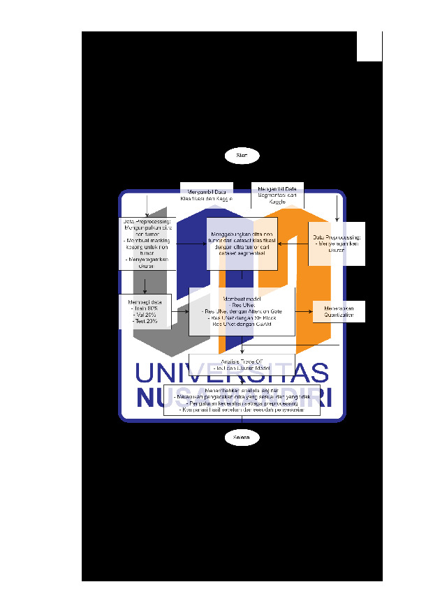
Segmentasi citra MRI tumor otak merupakan langkah penting dalam diagnosis dan perencanaan perawatan medis. Penelitian ini mengembangkan model Res-UNet dengan mekanisme attention, seperti attention gate (AG), SE block, dan CBAM, untuk mendeteksi segmentasi tumor otak. Evaluasi dilakukan menggunakan metrik Intersection over Union (IoU), dengan model terbaik mencapai IoU 0.845 setelah dynamic range quantization, ukuran model berkurang 75%, dan waktu inferensi 0.2055 detik per citra. Analisis menunjukkan bahwa tingkat kecerahan memengaruhi hasil segmentasi, di mana dengan perbaikan tingkat kecerahan meningkatkan akurasi pada citra dengan distribusi piksel homogen. Model ini dirancang untuk diterapkan pada perangkat keras dengan keterbatasan sumber daya, seperti di lingkungan rumah sakit. Hasil penelitian membuktikan bahwa Res-UNet dengan mekanisme attention dapat memberikan segmentasi yang akurat dan efisien, dengan potensi besar untuk mendukung diagnosis berbasis citra medis.
Unduhan


REFERENSI
[1] O. Ronneberger, P. Fischer, and T. Brox, “U-Net: Convolutional Networks for Biomedical Image Segmentation,” 2015, pp. 234–241. doi: 10.1007/978-3-319-24574-4_28.
[2] F. Isensee et al., “nnU-Net: Self-adapting Framework for U-Net-Based Medical Image Segmentation,” Informatik aktuell, p. 22, Sep. 2018, doi: 10.1007/978-3-658-25326-4_7.
[3] K. He, X. Zhang, S. Ren, and J. Sun, “Deep residual learning for image recognition,” Proceedings of the IEEE Computer Society Conference on Computer Vision and Pattern Recognition, vol. 2016-December, pp. 770–778, Dec. 2016, doi: 10.1109/CVPR.2016.90.
[4] O. Oktay et al., “Attention U-Net: Learning Where to Look for the Pancreas,” Apr. 2018, Accessed: Dec. 14, 2024. [Online]. Available: https://arxiv.org/abs/1804.03999v3
[5] J. Hu, L. Shen, and G. Sun, “Squeeze-and-Excitation Networks,” Proceedings of the IEEE Computer Society Conference on Computer Vision and Pattern Recognition, pp. 7132–7141, Dec. 2018, doi: 10.1109/CVPR.2018.00745.
[6] S. Woo, J. Park, J. Y. Lee, and I. S. Kweon, “CBAM: Convolutional block attention module,” Lecture Notes in Computer Science (including subseries Lecture Notes in Artificial Intelligence and Lecture Notes in Bioinformatics), vol. 11211 LNCS, pp. 3–19, 2018, doi: 10.1007/978-3-030-01234-2_1/TABLES/8.
[7] X. Li, Z. Fang, R. Zhao, and H. Mo, “Brain Tumor MRI Segmentation Method Based on Improved Res-UNet,” IEEE Journal of Radio Frequency Identification, vol. 8, pp. 652–657, 2024, doi: 10.1109/JRFID.2023.3349193.
[8] L. Huang, A. Miron, K. Hone, and Y. Li, “Segmenting Medical Images: From UNet to Res-UNet and nnUNet,” in 2024 IEEE 37th International Symposium on Computer-Based Medical Systems (CBMS), IEEE, Jun. 2024, pp. 483–489. doi: 10.1109/CBMS61543.2024.00086.
[9] X. Fang, H. Liu, G. Xie, Y. Zhang, and D. Liu, “Deep Neural Network Compression Method Based on Product Quantization,” in 2020 39th Chinese Control Conference (CCC), IEEE, Jul. 2020, pp. 7035–7040. doi: 10.23919/CCC50068.2020.9188698.
[10] J. Liu, L. Niu, Z. Yuan, D. Yang, X. Wang, and W. Liu, “PD-Quant: Post-Training Quantization Based on Prediction Difference Metric,” in 2023 IEEE/CVF Conference on Computer Vision and Pattern Recognition (CVPR), IEEE, Jun. 2023, pp. 24427–24437. doi: 10.1109/CVPR52729.2023.02340.
[11] J. Zhang, Z. Jiang, J. Dong, Y. Hou, and B. Liu, “Attention Gate ResU-Net for Automatic MRI Brain Tumor Segmentation,” IEEE Access, vol. 8, pp. 58533–58545, 2020, doi: 10.1109/ACCESS.2020.2983075.
[12] “Brain Tumor Segmentation.” Accessed: Dec. 22, 2024. [Online]. Available: https://www.kaggle.com/datasets/nikhilroxtomar/brain-tumor-segmentation
[13] “Brain Tumor MRI Dataset.” Accessed: Oct. 07, 2024. [Online]. Available: https://www.kaggle.com/datasets/masoudnickparvar/brain-tumor-mri-dataset/data
[14] S. F. Rabby, M. A. Arafat, and T. Hasan, “BT-Net: An end-to-end multi-task architecture for brain tumor classification, segmentation, and localization from MRI images,” Array, vol. 22, p. 100346, Jul. 2024, doi: 10.1016/j.array.2024.100346.
[15] S. Saifullah and R. Dreżewski, “Redefining brain tumor segmentation: a cutting-edge convolutional neural networks-transfer learning approach,” International Journal of Electrical and Computer Engineering (IJECE), vol. 14, no. 3, p. 2583, Jun. 2024, doi: 10.11591/ijece.v14i3.pp2583-2591.
[16] N. Musthafa, M. M. Masud, and Q. Memon, “Advancing Early-Stage Brain Tumor Detection with Segmentation by Modified_Unet,” in Proceedings of the 2024 8th International Conference on Medical and Health Informatics, New York, NY, USA: ACM, May 2024, pp. 52–57. doi: 10.1145/3673971.3674001.
[17] H. Alquran, M. Alslatie, A. Rababah, and W. A. Mustafa, “Improved Brain Tumor Segmentation in MR Images with a Modified U-Net,” Applied Sciences, vol. 14, no. 15, p. 6504, Jul. 2024, doi: 10.3390/app14156504.
[18] J. Wang, S.-Y. Lu, S.-H. Wang, and Y.-D. Zhang, “RanMerFormer: Randomized vision transformer with token merging for brain tumor classification,” Neurocomputing, vol. 573, p. 127216, Mar. 2024, doi: 10.1016/j.neucom.2023.127216.
[19] Md. M. Islam, P. Barua, M. Rahman, T. Ahammed, L. Akter, and J. Uddin, “Transfer learning architectures with fine-tuning for brain tumor classification using magnetic resonance imaging,” Healthcare Analytics, vol. 4, p. 100270, Dec. 2023, doi: 10.1016/j.health.2023.100270.
[20] S. G. De Benedictis, G. Gargano, and G. Settembre, “Enhanced MRI brain tumor detection and classification via topological data analysis and low-rank tensor decomposition,” Journal of Computational Mathematics and Data Science, vol. 13, p. 100103, Dec. 2024, doi: 10.1016/j.jcmds.2024.100103.
[21] A. M. Dikande Simo, A. Tchagna Kouanou, V. Monthe, M. Kameni Nana, and B. Moffo Lonla, “Introducing a deep learning method for brain tumor classification using MRI data towards better performance,” Inform Med Unlocked, vol. 44, p. 101423, 2024, doi: 10.1016/j.imu.2023.101423.
[22] G. D. Praveenkumar and R. Nagaraj, “Regularized Anisotropic Filtered Tanimoto Indexive Deep Multilayer Perceptive Neural Network learning for effective image classification,” Neuroscience Informatics, vol. 2, no. 2, p. 100063, Jun. 2022, doi: 10.1016/j.neuri.2022.100063.
[23] J. Peng and Y. Wang, “Medical Image Segmentation With Limited Supervision: A Review of Deep Network Models,” IEEE Access, vol. 9, pp. 36827–36851, 2021, doi: 10.1109/ACCESS.2021.3062380.
[24] N. Siddique, S. Paheding, C. P. Elkin, and V. Devabhaktuni, “U-Net and Its Variants for Medical Image Segmentation: A Review of Theory and Applications,” IEEE Access, vol. 9, pp. 82031–82057, 2021, doi: 10.1109/ACCESS.2021.3086020.
[25] “Brain MRI segmentation.” Accessed: Oct. 07, 2024. [Online]. Available: https://www.kaggle.com/datasets/mateuszbuda/lgg-mri-segmentation/data
[26] N. Ibtehaz and M. S. Rahman, “MultiResUNet : Rethinking the U-Net architecture for multimodal biomedical image segmentation,” Neural Networks, vol. 121, pp. 74–87, Jan. 2020, doi: 10.1016/j.neunet.2019.08.025.
[27] H. Lu, Y. She, J. Tie, and S. Xu, “Half-UNet: A Simplified U-Net Architecture for Medical Image Segmentation,” Front Neuroinform, vol. 16, Jun. 2022, doi: 10.3389/fninf.2022.911679.
[28] A. Tiwari, S. Srivastava, and M. Pant, “Brain tumor segmentation and classification from magnetic resonance images: Review of selected methods from 2014 to 2019,” Pattern Recognit Lett, vol. 131, pp. 244–260, Mar. 2020, doi: 10.1016/j.patrec.2019.11.020.
[29] F. Milletari, N. Navab, and S. A. Ahmadi, “V-Net: Fully convolutional neural networks for volumetric medical image segmentation,” Proceedings - 2016 4th International Conference on 3D Vision, 3DV 2016, pp. 565–571, Dec. 2016, doi: 10.1109/3DV.2016.79.
[30] A. Vaswani et al., “Attention Is All You Need,” Adv Neural Inf Process Syst, vol. 2017-December, pp. 5999–6009, Jun. 2017, Accessed: Dec. 14, 2024. [Online]. Available: https://arxiv.org/abs/1706.03762v7
[31] A. F. M. M. Rahman and Md. A. Hossain, “Attention-refined U-Net with Skip Connections for Effective Brain Tumor Segmentation from MRI Images,” in 2023 26th International Conference on Computer and Information Technology (ICCIT), IEEE, Dec. 2023, pp. 1–6. doi: 10.1109/ICCIT60459.2023.10441092.
[32] T. Li, J. J. Liu, Y. Tai, and Y. Tian, “Brain tumor segmentation with attention-based U-Net,” in Second IYSF Academic Symposium on Artificial Intelligence and Computer Engineering, W. Qin, Ed., SPIE, Dec. 2021, p. 87. doi: 10.1117/12.2623112.
[33] Y.-Z. Fang and J.-D. Huang, “Enhancing Brain Tumor Segmentation with Deep Supervision and Attention Mechanisms: Advances in the nnU-Net Framework,” in 2024 IEEE International Symposium on Biomedical Imaging (ISBI), IEEE, May 2024, pp. 1–4. doi: 10.1109/ISBI56570.2024.10635180.
[34] M. AskariHemmat et al., “U-Net Fixed-Point Quantization for Medical Image Segmentation,” 2019, pp. 115–124. doi: 10.1007/978-3-030-33642-4_13.
[35] A. Raju and N. Sinha, “SSEGEP: Small Segment Emphasized Performance Evaluation Metric for Medical Image Segmentation,” JOURNAL OF MACHINE LEARNING IN FUNDAMENTAL SCIENCES, vol. 2022, no. 1, 2022, doi: 10.31526/JMLFS.2022.229.
[36] Z. Wang, Y. Zou, and P. X. Liu, “Hybrid dilation and attention residual U-Net for medical image segmentation,” Comput Biol Med, vol. 134, p. 104449, Jul. 2021, doi: 10.1016/j.compbiomed.2021.104449.
[37] X. Gan, L. Wang, Q. Chen, Y. Ge, and S. Duan, “GAU-Net: U-Net Based on Global Attention Mechanism for brain tumor segmentation,” J Phys Conf Ser, vol. 1861, no. 1, p. 012041, Mar. 2021, doi: 10.1088/1742-6596/1861/1/012041.
[38] R. Li, S. Zheng, C. Duan, J. Su, and C. Zhang, “Multistage Attention ResU-Net for Semantic Segmentation of Fine-Resolution Remote Sensing Images,” IEEE Geoscience and Remote Sensing Letters, vol. 19, pp. 1–5, 2022, doi: 10.1109/LGRS.2021.3063381.
[39] J. Jiang, E. Sharif, H. Um, S. Berry, and H. Veeraraghavan, “Local block-wise self attention for normal organ segmentation,” Sep. 2019, Accessed: Dec. 14, 2024. [Online]. Available: https://arxiv.org/abs/1909.05054v1
[40] D. Zhu, “Attention-based U-Net Denoising Network,” in 2023 IEEE International Conference on Sensors, Electronics and Computer Engineering (ICSECE), IEEE, Aug. 2023, pp. 746–750. doi: 10.1109/ICSECE58870.2023.10263450.
[41] W. Y. Chung, I. H. Lee, and C. G. Park, “Lightweight Infrared Small Target Detection Network Using Full-Scale Skip Connection U-Net,” IEEE Geoscience and Remote Sensing Letters, vol. 20, pp. 1–5, 2023, doi: 10.1109/LGRS.2023.3276326.
[42] L. Dongdong, T. Wenjie, L. Songlin, and Q. Xiaolan, “Improved S2ANet based on attention mechanism for small target detection in remote sensing images,” in 2021 CIE International Conference on Radar (Radar), IEEE, Dec. 2021, pp. 942–945. doi: 10.1109/Radar53847.2021.10028564.
[43] W. Guo, Q. Feng, X. Li, S. Yang, and J. Yang, “Grape leaf disease detection based on attention mechanisms,” International Journal of Agricultural and Biological Engineering, vol. 15, no. 5, pp. 205–212, 2022, doi: 10.25165/j.ijabe.20221505.7548.
[44] N. S. Punn and S. Agarwal, “RCA-IUnet: a residual cross-spatial attention-guided inception U-Net model for tumor segmentation in breast ultrasound imaging,” Mach Vis Appl, vol. 33, no. 2, p. 27, Mar. 2022, doi: 10.1007/s00138-022-01280-3.
[45] K. Yamamoto, “Learnable Companding Quantization for Accurate Low-bit Neural Networks,” in 2021 IEEE/CVF Conference on Computer Vision and Pattern Recognition (CVPR), IEEE, Jun. 2021, pp. 5027–5036. doi: 10.1109/CVPR46437.2021.00499.
[46] Y. Jung, H. Kim, Y. Choi, and L.-S. Kim, “Quantization-Error-Robust Deep Neural Network for Embedded Accelerators,” IEEE Transactions on Circuits and Systems II: Express Briefs, vol. 69, no. 2, pp. 609–613, Feb. 2022, doi: 10.1109/TCSII.2021.3103192.
[47] Q. Jin, L. Yang, and Z. Liao, “Towards Efficient Training for Neural Network Quantization,” Dec. 2019, Accessed: Dec. 14, 2024. [Online]. Available: https://arxiv.org/abs/1912.10207v1
[48] S. Dai, R. Venkatesan, H. Ren, B. Zimmer, W. J. Dally, and B. Khailany, “VS-Quant: Per-vector Scaled Quantization for Accurate Low-Precision Neural Network Inference,” Feb. 2021, Accessed: Dec. 15, 2024. [Online]. Available: https://arxiv.org/abs/2102.04503v1
[49] X. Wangl, Y. Zhong, and J. Dong, “A New Low-Bit Quantization Algorithm for Neural Networks,” Chinese Control Conference, CCC, vol. 2023-July, pp. 8509–8514, 2023, doi: 10.23919/CCC58697.2023.10241028.
[50] J. Faraone, N. Fraser, M. Blott, and P. H. W. Leong, “SYQ: Learning Symmetric Quantization for Efficient Deep Neural Networks,” Proceedings of the IEEE Computer Society Conference on Computer Vision and Pattern Recognition, pp. 4300–4309, Dec. 2018, doi: 10.1109/CVPR.2018.00452.
[51] P. Nayak, D. Zhang, and S. Chai, “Bit Efficient Quantization for Deep Neural Networks,” Proceedings - 5th Workshop on Energy Efficient Machine Learning and Cognitive Computing, EMC2-NIPS 2019, pp. 52–56, Dec. 2019, doi: 10.1109/EMC2-NIPS53020.2019.00020.
[52] E. Park, J. Ahn, and S. Yoo, “Weighted-entropy-based quantization for deep neural networks,” Proceedings - 30th IEEE Conference on Computer Vision and Pattern Recognition, CVPR 2017, vol. 2017-January, pp. 7197–7205, Nov. 2017, doi: 10.1109/CVPR.2017.761.
[53] D. Liu, X. Chen, C. Ma, and X. Liu, “Hyperspherical Quantization: Toward Smaller and More Accurate Models,” Proceedings - 2023 IEEE Winter Conference on Applications of Computer Vision, WACV 2023, pp. 5251–5261, 2023, doi: 10.1109/WACV56688.2023.00523.
[54] Z. Dong, Z. Yao, D. Arfeen, A. Gholami, M. W. Mahoney, and K. Keutzer, “HAWQ-V2: Hessian Aware trace-Weighted Quantization of Neural Networks,” Adv Neural Inf Process Syst, vol. 2020-December, Nov. 2019, Accessed: Dec. 15, 2024. [Online]. Available: https://arxiv.org/abs/1911.03852v1
[55] P.-F. Yan et al., “Accuracy of conventional MRI for preoperative diagnosis of intracranial tumors: A retrospective cohort study of 762 cases,” International Journal of Surgery, vol. 36, pp. 109–117, Dec. 2016, doi: 10.1016/j.ijsu.2016.10.023.
[56] G. Mahesh and Y. K. M, “Brain Tumor Detection and Classification Using MRI Images,” Int J Res Appl Sci Eng Technol, vol. 12, no. 10, pp. 856–863, Oct. 2024, doi: 10.22214/ijraset.2024.64719.
[57] K. R. Laukamp et al., “Fully automated detection and segmentation of meningiomas using deep learning on routine multiparametric MRI,” Eur Radiol, vol. 29, no. 1, pp. 124–132, Jan. 2019, doi: 10.1007/s00330-018-5595-8.
[58] D. Sreedhar and A. Fathi Kazerooni, “Evaluating the clinical applicability of neural networks for meningioma tumor segmentation on 3D MRI,” J Emerg Investig, 2024, doi: 10.59720/23-265.
[59] A. B. Abdusalomov, M. Mukhiddinov, and T. K. Whangbo, “Brain Tumor Detection Based on Deep Learning Approaches and Magnetic Resonance Imaging,” Cancers (Basel), vol. 15, no. 16, p. 4172, Aug. 2023, doi: 10.3390/cancers15164172.
[60] A. S. Musallam, A. S. Sherif, and M. K. Hussein, “A New Convolutional Neural Network Architecture for Automatic Detection of Brain Tumors in Magnetic Resonance Imaging Images,” IEEE Access, vol. 10, pp. 2775–2782, 2022, doi: 10.1109/ACCESS.2022.3140289.
[61] N. Ullah et al., “An Effective Approach to Detect and Identify Brain Tumors Using Transfer Learning,” Applied Sciences, vol. 12, no. 11, p. 5645, Jun. 2022, doi: 10.3390/app12115645.
Kiriman Terbaru
-

08 Jul 2026
-
08 Jul 2026
-

08 Jul 2026
-

08 Jul 2026
-

08 Jul 2026